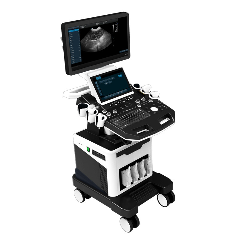
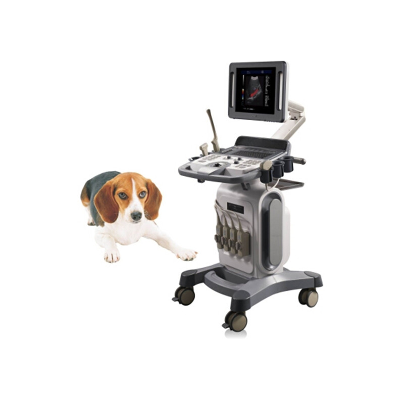
product description
Features
- Operating system: Windows 8 operating system
- Pulse Doppler Imaging (PW)
- Direction Power Doppler Imaging (DPDI)
- B/C/D Real-time Three Synchronous Imaging
- Compound Imaging
- Tissue Harmonic Imaging (THI)
- 2B/4B Imaging Modes
- System language option: Chinese, English, French, Russian, Spanish
- Main monitor: ≥5 inch
Technical Specifications
|
PARAMETER |
SPECIFICATION |
|
2D Imaging Mode |
|
|
Gain |
0-100, Step 1 adjustable |
|
TGC |
8 segment adjustable |
|
Dynamic |
20-280, 20 levels adjustable |
|
Pseudo color |
0-11, adjustable |
|
Sound power |
5%-100%, step 5% adjustable |
|
Body mark |
≥18 kinds optional |
|
Maximum focus |
≥6, which can be moved throughout the whole process |
|
Grey scale map |
0-7, 7 levels adjustable |
|
Filter |
0-4 |
|
Scanning range |
50%-100% |
|
Frame correlation |
0-4, 4 level adjustable |
|
Line density |
low, middle, high, 3 levels adjustable |
|
Noise reduction |
0-14, 4 level adjustable |
|
The screen has real-time display of sound power, probe frequency, dynamic range, pseudo color, gray scale and other 14 parameters can be adjusted. |
|
|
Color Doppler Imaging Mode |
|
|
Color Frame correlation |
0-12, 12 levels adjustable |
|
Color map |
0-7, 7 levels adjustable |
|
Color flip |
adjustable |
|
Base line |
11 levels, adjustable |
|
Line density |
low, high, 2 levels adjustable |
|
Filter |
0-5 levels adjustable |
|
B / C real-time split screen mode |
|
|
Spectral Doppler Imaging Mode |
|
|
Sampling volume angle correction |
-80°~80°adjustable |
|
Sample volume |
0.5mm-20mm adjustable |
|
Frequency |
2.5MHz, 3.0MHz |
|
Base line |
11 levels, adjustable |
|
Pseudo color |
0-5 |
|
Parameter display |
≥4 kinds, adjustable |
|
Speed scale |
32.8-328cm/s (different probes have different ranges) |
|
Spectrum envelope function |
real time automatic spectrum envelope, manual spectrum envelope, and other. The system automatically analyses and displays various data such as PS, ED, RI, PI, S/D, HR, etc. |
|
Grey map |
0-7 |
|
Filter |
0-8 |
|
Dynamic range |
10-95, step 5 |
|
Noise reduction |
0-28 |
|
Sound volume |
0-100 |
|
3D Imaging (optional) |
|
|
Fast angle |
supports 0°, 90°, 180°, 270° rotation for 3D View |
|
Display mode |
one image, two images, four images |
|
Reconstruction mode |
Real Skin, Surface, Max, Min, X Rax |
|
Pseudo color |
0-7, 7 levels adjustable |
|
Zoom |
5 levels |
|
Contrast |
0%-100% |
|
Threshold level |
0%-100% |
|
Smooth |
≥3 levels |
|
Image rotation |
X/Y/Z Axis |
|
Brightness |
0%-100% |
|
4D Imaging (optional) |
|
|
Fast angle |
supports 0°, 90°, 180°, 270° rotation for 3D View |
|
Display model |
one image, two images, four images |
|
Reconstruction mode |
Real Skin, Surface, Max, Min, X Rax |
|
Pseudo color |
0-7, 7 levels adjustable |
|
Zoom |
5 levels |
|
Contrast |
0%-100% |
|
Threshold level |
0%-100% |
|
Smooth |
≥3 levels, adjustable |
|
Image rotation |
X/Y/Z Axis |
|
Line density |
2 levels, adjustable |
|
Measurement and Analysis |
|
|
General measurement |
distance, area, angle, time, slope, heart rate, speed, acceleration, blood flow path, blood flow spectrum trace, resistance index/pulsation index, etc |
|
OB measurement data |
Cannie, Feline, Bovine, Ovine, Equine |
|
Measurement line |
color and type can be adjusted at will (including the activation color and the completion color) |
|
Measurement result |
display position and font size can be adjusted as needed |
|
Professional data package |
Abdomen, OB, Urology, etc |
Various Probes
- Convex probe (detecting depth: 30-255mm)
Fundamental Frequency: 2.5MHz/ 3.0MHz/ 3.5MHz/ 4.0MHz
Harmonic Frequency: H4.0MHz/ H5.0MHz
- Linear probe (detecting depth: 20-128mm)
Fundamental Frequency: 6.0MHz/ 7.5MHz/ 8.5MHz/ 10.0MHz
Harmonic Frequency: H10.0MHz
- Rectal probe (detecting depth: 30-156mm)
Fundamental Frequency: 4.5MHz/ 6.0MHz/ 7.0MHz/ 9.0MHz
Harmonic Frequency: H8.0MHz
- Phased array probe (detecting depth: 100-244mm)
Fundamental Frequency: 2.5MHz/ 3.0MHz/ 3.5MHz/ 4.0MHz
Harmonic Frequency: H3.0MHz/ H4.0MHz
- 4D volume probe (detecting depth: 30-237mm)
Fundamental Frequency: 2.0MHz/ 3.0MHz/ 4.5MHz/ 6.0MHz
Harmonic Frequency: H5.0MHz
- Micro-convex probe R15 (detecting depth: 30-111mm)
Fundamental Frequency: 4.0MHz/ 6.0MHz/ 7.0MHz/ 8.0MHz
Harmonic Frequency: H8.0MHz
- Micro-convex probe R11 (detecting depth: 30-111mm)
Fundamental Frequency: 4.5MHz/ 6.0MHz/ 7.0MHz/ 9.0MHz
Harmonic Frequency: H8.0MHz
- Micro-convex probe R20 (detecting depth: 30-111mm)
Fundamental Frequency: 4.5MHz/ 6.0MHz/ 7.0MHz/ 9.0MHz
Harmonic Frequency: H8.0MHz
Graphic and Text Management System
- Host build in 128G hard disk, start fast and stable.
- Movie playback: ≥600 frames.
- Report type: ≥6.
- One key fast report graphic and text management.
- Image format: BMP, DCM, JPG.
- Internal file information management system: can record patient number, name, check number, check date and so on, and can be searched and managed by number, check number, name and so on.
Configuration
- Full Digital Color Doppler Ultrasound Diagnostic System: 1
- Video printer (optional), Laser printer (optional), Trolley


